
Ophthalmology Learning Module
Rudolf Riester GmbH
Ophthalmology product line
Last edited: Feb. 16, 2021
Ophthalmology is the branch of medicine that deals with the anatomy, physiology and diseases of the eye and the visual system. With the aging global population, eye health is becoming increasingly important due to more incidences of age-related eye diseases. Cataracts are the main cause of vision impairment worldwide and represent the second most common surgery performed in England. Most eye diseases can be successfully treated if caught early and can be managed effectively with existing treatments and medicines.
The eye is the organ which enables us to see and interpret the shapes, colours, and dimensions of elements of our surroundings by processing the light they reflect or emit.

Eyesight
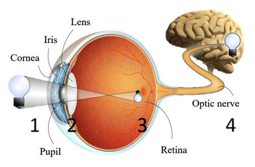
How do we see?
1) Light waves from an object come in through the cornea where they are first converged before reaching the pupil, the circular opening in the centre of the coloured iris. The size of the pupil depends on the intensity of the incoming light. In a dark environment, the pupil dilates (enlarges) to allow in as much light as possible. The opposite happens in a bright environment: the pupil constricts.
2) A crystalline lens sits behind the iris and the pupil and further converges the light waves, forming a reversed and inverted image on the retina.
3) As they penetrate the various layers of the retina, light waves are converted to electrical signals which travel through the optic nerve along the visual pathway until it reaches the occipital cortex at the back of the brain.
4) This is where the brain interprets the electrical signal into an image that it is able to visualize.
Refractive errors
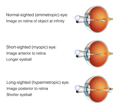
The brain is not able to form a clear image if the light waves are not focused on the retina. A short-sighted (or near-sighted, myopic) eye for example would have its image formed anterior to the retina due to the eyeball being long lengthwise. In other words, an object at a distance would appear blurry, a condition also known as myopia. A long-sighted (or far-sighted, hypermetropic) eye would have it formed posterior to the retina due to the eyeball being short. Close objects would appear blurry in this condition also known as presbyopia. Such problems are known as refractive errors.
Eye tests and eye screening
Depending on age, ethnicity and health conditions, biannually, annually or biennially eye tests are recommended. In addition to those, some countries have diabetic eye screening programmes in place to detect diabetic retinopathy at an early stage. In the UK for example, “from the age of 12, all people with diabetes are offered an annual diabetic eye test to check for early signs of diabetic retinopathy.” (NHS, 2017) This is motivated by the following facts:
-
Diabetic retinopathy is the leading cause of preventable blindness in the 20-74 years old population in western countries.
-
Diabetes diagnoses are growing exponentially around the world
- Almost all patients with diabetes type 1 and > 60% of diabetes type 2 who have had the disease for over 20 years develop retinopathy.
- Retinopathy can generally be treated if detected early. If the retina is not severely damaged, even patients presenting with an advanced retinopathy have a 95% chance of keeping their vision when they get treatment.
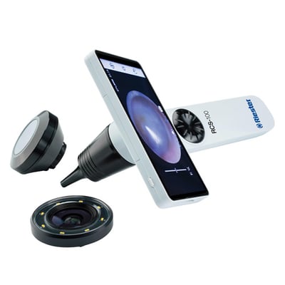
The International Council of Ophthalmology recommends that a retinal examination be performed either by direct or indirect ophthalmoscopy or slit-lamp biomicroscopy of the retina, or by “retinal (fundus) photography with any of the following: 30-degree to wide field, mono or stereo photography, and dilated or undilated photography” (Vujosevic et al., 2020) . For the latter, telemedicine is a suitable approach.
Telemedicine
A study was conducted by Malerbi et al. (Diabetology & Metabolic Syndrome, 2015) on a large and heterogenous type 1 diabetes population in Brazil to compare binocular indirect ophthalmoscopy to telemedicine protocols of digital retinography. They found a substantial agreement between the two modalities when the pupil was dilated (mydriasis) and therefore recommended the use of telemedicine for digital retinography (mydriatic, preferably with one field centred on the fovea and the other field centred on the optic disc) in developing countries. On the other hand, they argued against non-mydriatic retinography due to a significant loss of information in the images obtained.
Ophthalmoscopy
Ophthalmoscopy, also called funduscopy, is a test that allows an examiner to see inside the fundus of the eye and other structures to determine the health of the retina, vitreous humor and optic disc and using an ophthalmoscope. The ophthalmoscope is the most frequently used instrument for any optician, optometrist or ophthalmologist as it can uncover a wide variety of conditions. A patient complaining of headache might have swollen discs, cupped discs are often seen in glaucoma patients and increased intracranial and arterial hypertension can indicate serious conditions.

There are two main types of ophthalmoscopes:
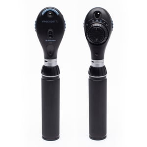
Direct ophthalmoscope: handheld instrument that consists of an eye piece, a light source, a concave mirror, a set of lenses and a handle containing batteries. Most commonly used during routine examinations. The image of the fundus can be magnified up to 15x and is rendered upright but it is difficult to visualize the peripheral retina using a direct ophthalmoscope. No 3D rendition of the interior of the eye is available. Click here to learn more about direct ophthalmoscopes and their working principles.
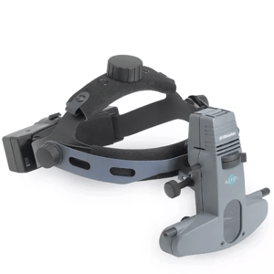
Indirect ophthalmoscope: comes in monocular or binocular form and produces an inverted image. In both types, the field of view is wider than with the direct ophthalmoscope but the magnification is lower. With the binocular version, a 3D rendition of the interior of the eye is obtained which allows for a more thorough examination and therefore makes it a more popular choice. The peripheral retina can be viewed. Most commonly used for patients presenting with an eye condition or certain complexities. Typically consists of a headband, binocular lens with mirrors and a light source.
Retinoscopy
Retinoscopy is a procedure used to measure how well one can see objects at varying distance and determine the refractive error so that vision correction can be adapted. Its purpose is to inspect the light moving across the fundus. It is an indispensable test for paediatric patients and can prove very helpful with non-communicative and non-cooperative patients. Using a retinoscope, the examiner can determine the correct prescription for eye glasses or lenses without needing the patient’s feedback.
Basic principle of retinoscopy
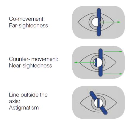
By sweeping the beam across the pupil and observing how the light rays are reflected off the patient’s retina, the examiner can measure the extent of refractive error in the eyes of the patient. If the light reflected back is moving in synchrony with the beam, in the same direction, we have a “with-reflex”, indicative of far-sightedness due to the light focusing behind the eye. When a lens with the correct prescription is placed in front of the eye, the beam hits the retina perfectly: the light is focused on the retina and the reflex is neutralised (no movement). When the light reflected back from the retina is moving in the opposite direction from the beam (counter-movement), we have an “against-reflex”, indicative of near-sightedness due to the light focusing in front of the retina.
Retinoscope
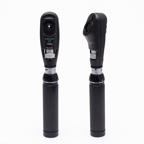
Retinoscopy is performed using a handheld instrument called a retinoscope that comprises:
-
an eyepiece with a mirror with a central hole and a variable condensing lens
-
a light source in the form of a spot or streak bulb
-
a collar that can move up and down to change the vergence of the light or rotate to change the angle of the beam
-
an on/off/brightness control
Modern retinoscopes are self-illuminated and are categorized as streak or spot retinoscopes, depending on their light source. Both project an intercept (a streak of light) but only the streak retinoscope has the ability to rotate the intercept which makes it the preferred instrument in modern ophthalmology.
Streak retinoscope
Reflects a beam of light from a linear light source.
-
Operates with plane mirror effect (plano mode) or concave mirror effect
- Ideal to detect astigmatism
- More commonly used as offers dual modes
Spot retinoscope
Reflects a beam of light from a circular light source.
-
Operates with plane mirror effect
-
More visual information available (optical phenomena that take place away from the center, subtle changes in brightness, colour or cylinder…)
This video created by Dr. Tim Root from OphthoBook brilliantly explains, in a simplified manner, the working principle of the retinoscope.
Light source
Ophthalmoscopes and retinoscopes come with either a filament bulb relying on a gas (halogen or xenon) or an LED bulb relying on diodes and electrons.
Halogen bulbs are the least efficient in terms of lighting, lifetime and heat release, but they are usually the cheapest. Xenon bulbs last longer, produce a brighter and whiter light (the halogen is more yellowish; the xenon, blueish) and release less heat. LED bulbs offer similar improvements as the xenon bulbs but take them one step further – while LEDs typically shine less bright than xenon light, they produce a more accurate pigmentation, stay cool to touch and last exponentially longer, making them cost effective despite the higher cost upfront.
-
Lighting: filament vs LED
An accurate pigmentation is made possible by the use of LED light due to its natural temperature colour. LED also provides more consistent light throughout the whole area being viewed than halogen. While xenon has long been favoured for high-end medical lighting applications due to its bright, white light, LED bulbs can now produce a similar level of bright, white light.
-
Lifetime: filament vs LED
An LED bulb can last between 50,000-100,000 hours, more than 10x longer than an average filament bulb. This is 6-12 years of continuous use before needing replacement.
-
Heat release: filament vs LED
Filament bulbs produce a lot of heat (risk of burns or fire). LED bulbs are cool to the touch which makes them safer.
-
Cost: filament vs LED
Filament bulbs are cheaper to replace than LED bulbs. LED bulbs have a higher cost upfront but last exponentially longer and require less maintenance, making them a cheaper alternative over the long run.

Did you know?
Works Cited
Damery, E. Lescanne, K. Reffet, C. Aussedat, D. Bakhos, Interest of video-otoscopy for the general practitioner, European Annals of Otorhinolaryngology, Head and Neck Diseases, Volume 136, Issue 1, 2019, Pages 13-17, ISSN 1879-7296, https://doi.org/10.1016/j.anorl.2018.10.016.
How Do We Hear? | NIDCD (nih.gov)
Kristen L. Yancey, Loyce J. Cheromei, Jackie Muhando, Jim Reppart, James L. Netterville, Asitha D.L. Jayawardena, Pediatric hearing screening in low-resource settings: Incorporation of video-otoscopy and an electronic medical record, International Journal of Pediatric Otorhinolaryngology, Volume 126,2019, 109633, ISSN 0165-5876, https://doi.org/10.1016/j.ijporl.2019.109633.
Malene Nøhr Demant, Ramon Gordon Jensen, Mahmood F. Bhutta, Gunnar Hellmund Laier, Jørgen Lous, Preben Homøe, Smartphone otoscopy by non-specialist health workers in rural Greenland: A cross-sectional study, International Journal of Pediatric Otorhinolaryngology, Volume 126, 2019, 109628, ISSN 0165-5876, https://doi.org/10.1016/j.ijporl.2019.109628.
Manan Udayan Shah, Maheep Sohal, Tulio A. Valdez, Christopher R. Grindle, iPhone otoscopes: Currently available, but reliable for tele-otoscopy in the hands of parents?, International Journal of Pediatric Otorhinolaryngology, Volume 106, 2018,Pages 59-63, ISSN 0165-5876, https://doi.org/10.1016/j.ijporl.2018.01.003.
Modupe Oyewumi, Michael G. Brandt, Brian Carrillo, Adelle Atkinson, Karl Iglar, Vito Forte, Paolo Campisi, Objective Evaluation of Otoscopy Skills Among Family and Community Medicine, Pediatric, and Otolaryngology Residents, Journal of Surgical Education, Volume 73, Issue 1,2016, Pages 129-135, ISSN 1931-7204, https://doi.org/10.1016/j.jsurg.2015.07.011.
Pneumatic Otoscope Examination: Overview, Indications, Contraindications (medscape.com)
Thorbjörn Lundberg, Leigh Biagio de Jager, De Wet Swanepoel, Claude Laurent, Diagnostic accuracy of a general practitioner with video-otoscopy collected by a health care facilitator compared to traditional otoscopy, International Journal of Pediatric Otorhinolaryngology, Volume 99,2017, Pages 49-53, ISSN 0165-5876, https://doi.org/10.1016/j.ijporl.2017.04.045.