
Dermoscopy, also known as dermatoscopy, is a non-invasive diagnostic technique used worldwide for mole screening and skin cancer diagnosis. A large meta-analysis by Kittler et al. (The Lancet Oncology, 2002) showed that the accuracy of melanoma diagnosis was increased by about 50% when using a dermoscope in comparison to the unaided eye. The dermoscope allowed a greater visualisation of structures in the skin which were otherwise invisible to the naked eye, thus improving physicians’ confidence in their clinical diagnosis. However, the study also found a strong correlation between the level of accuracy and the degree of experience of the examiner. In other words, an examiner without proper training using a dermoscope did not achieve a better accuracy than an examiner making a diagnosis with their naked eye.
The principles of dermoscopy have been known for over 300 years but the rate of utilization has only recently started to increase in a significantly manner. Europe benefited from a head start, having conducted rigorous studies in the 1980s and 1990s to establish dermoscopic/histologic correlates and algorithms for melanoma diagnosis.
United States
Australia
France
United Kingdom
India
Basic principle
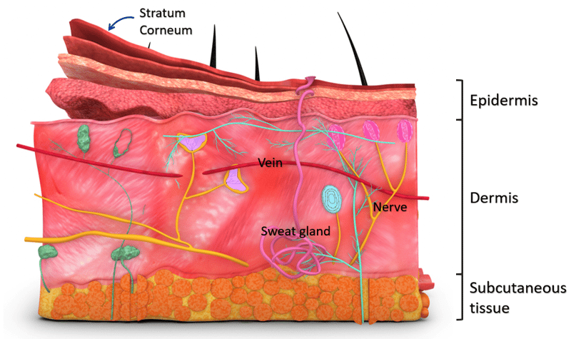
The skin is made up of several layers of various composition which makes it challenging for the naked eye to see past the outermost layer (the stratum corneum).
Light must travel through air until it reaches the skin before some of it is reflected back to the eye to give our brain visual information. This reflection is made possible by the difference in refractive indices of the media light travels through. Generally, some fraction of the light is reflected from the interface, and the remainder is refracted.
The fraction of light reflected between two mediums is predicted by the following equation:

Where n1 is the refractive index of medium 1 and n2 is the refractive index of medium 2. From that equation, if we had n1 ≈ n2, then light would be able to travel between the two mediums without being reflected because R → 0. (Remember that R is the fraction of light reflected between two mediums.)
Thinking back about the skin, this ideal scenario would allow us to see past the first layer because the light would in fact reach deeper before being reflected. In reality, when looking at the skin with a naked eye, this can’t happen because light encounters both air (n = 1.00) and the stratum corneum (n = 1.55). This mismatch in refraction index causes a reflected ‘glare’ (specular reflectance) which carries little visual information about underlying structures.
We want to reduce glare to obtain visual information about underlying structures. This can be achieved in 2 ways:
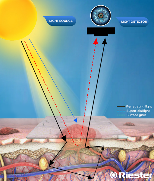
By reducing the refractive index mismatch between air and the stratum corneum, for example by adding a glass (n = 1.50) and an interface liquid (n between 1.50 and 1.55). This is the basic principle behind non-polarised dermoscopy (NPD). In the illustration on the left, the dermoscope's light source is shown in the top left corner. It irradiates rays of light in the form of the black, blue and red arrows. The blue arrow represents surface glare, here eliminated by the use of the immersion fluid (commonly used: mineral oil, ultrasound gel, 70% alcohol). The red and black arrows provide visual information on the skin lesion by penetrating skin layers: the red arrows only superficially, within the epidermis, but the black arrow undergoes scattering event through deeper layers and thus returns a different visual information to the dermoscope's light detector. However, when using a glass and an interface liquid, there are only few scattering events and most of the light returning to the detector is superficial. Thus, superficial light (red arrow) is the main source of contrast when using non-polarised dermoscopy which means that NPD it ideal for observing structures located between the stratum corneum and superficial papillary dermis
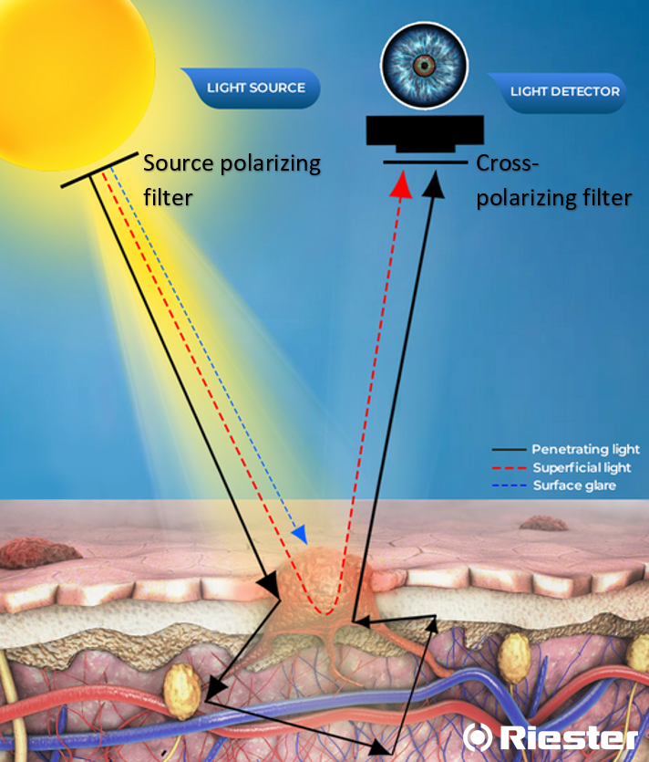
By polarising the incident light. This is achieved using filters and it allows visualization of structures deeper than what we can achieve simply by reducing the refractive index mismatch. It is referred to as polarised dermoscopy (PD). The light emitted by the dermoscope passes through a polarizer, generating polarized (unidirectional) light. It continues to the skin where it travels through various layers and gets scattered before a fraction of the light gets reflected back to the dermoscope (its light detector, more specifically). The cross-polarizing filter placed before the light detector only allows specific photons through, and to obtain such photos, enough scattering events must have happened. The deeper light travels, the higher the likelihood of scattering events.
Using the NPD's reference to black, blue and red arrows, it would compare as follows: in both cases, the blue arrow is eliminated (no surface glare); the proportion of red arrows reaching the detector is much higher than black arrows in NPD; the proportion of black arrows reaching the detector is much higher in PD. This means that what we see when using a polarised dermoscope is a reflection of deeper structures than we would see using a non-polarised dermoscope.
Types of dermoscope
Dermoscopes can be non-polarised (NPD) or polarised (PD). Hybrid devices are available as well, combining both types and making it possible to use with or without a liquid interface.
It is important to note that NPDs and PDs complement each other in their capabilities without being equivalent.
Non-polarised dermoscope (NPD)
With a long history of use, these devices, also known as contact oil immersion dermoscopes, are the most established. Recent studies comparing them to newer technologies involving polarised light have found NPDs to be better for blue-white colours, peppering, milia-like cysts and comedo-like openings.
NPD involves the use of a liquid interface (ideally with a refraction index equal to that of the skin, 1.55) and a glass plate mounted on the dermoscope (refraction index approximately 1.52). The liquid interface is used as an optical link between the stratum corneum and the glass plate. This is based on the principle that light passing through media with similar refractive indexes tends to maintain its propagation direction. With this method, there is less light reflection and refraction on the skin surface and deeper structures can thus be visualized when compared to the naked eye.
Polarised dermoscope
The polarised dermoscope (PD) makes use of two in-built filters: one is positioned between the skin and the light source and the other is between the skin and the light detector. The light passing through the first filter will reach the skin: some of it will get reflected and some will penetrate deeper levels and get backscattered. The second filter will only allow the backscattered light (no longer polarized) to reach the detector and thus the eye. That way, only the backscattered light from deeper levels of the skin is ultimately captured and the superficially reflected light which is normally responsible for the glistening aspect of the skin is avoided. It is not necessary to use a glass plate and a liquid interface with the polarised dermoscope.
PD is better at showing white shiny structures, pinks/reds, variable pigmentation and dermal vessels.
Because NPDs were traditionally used in dermoscopy training, training material usually showcases images captured without the use of polarised light. Special attention needs to be taken when using PDs not to refer to the traditional diagnostic criteria as certain shades such as blue and brown are rendered differently when compared to NPDs.
Works Cited
Amy Lake & Bolette Jones (2015) Dermoscopy: to cross-polarize, or not to cross-polarize, that is the question, Journal of Visual Communication in Medicine, 38:1-2, 36-50, DOI: 10.3109/17453054.2015.1046371
Benvenuto-Andrade C, Dusza SW, Agero ALC, et al. Differences Between Polarized Light Dermoscopy and Immersion Contact Dermoscopy for the Evaluation of Skin Lesions. Arch Dermatol. 2007;143(3):329–338. doi:10.1001/archderm.143.3.329
Butler, Thomas & Matin, Rubeta & Affleck, Andrew & Fleming, Colin & Bowling, Jonathan. (2015). Trends in dermoscopy use in the UK: results from surveys in 2003 and 2012. Dermatology practical & conceptual. 5. 29-38. 10.5826/dpc.0502a04.
Chen LL, Wei EX, Ma F, Keri J, Hu S. Rates of Dermoscopy Use for Melanoma Diagnosis in the Miami VA Medical Center. JAMA Dermatol. 2017;153(6):602–603. doi:10.1001/jamadermatol.2016.6025
Dermoscopy – an overview | Primary Care Dermatology Society | UK (pcds.org.uk)
Dermoscopy: Looking skin deep (practice) | Khan Academy
Estee L. Psaty, Allan C. Halpern,Current and emerging technologies in melanoma diagnosis: the state of the art,Clinics in Dermatology,Volume 27, Issue 1,2009,Pages 35-45,ISSN 0738-081X,https://doi.org/10.1016/j.clindermatol.2008.09.004.
H Kittler, H Pehamberger, K Wolff, M Binder, Diagnostic accuracy of dermoscopy, The Lancet Oncology, Volume 3, Issue 3, 2002, Pages 159-165, ISSN 1470-2045, https://doi.org/10.1016/S1470-2045(02)00679-4
Moulin C, Poulalhon N, Duru G, Debarbieux S, Dalle S, Thomas L. Dermoscopy use by French private practice dermatologists: a nationwide survey. Br J Dermatol. 2013 Jan;168(1):74-9. doi: 10.1111/j.1365-2133.2012.11216.x. Epub 2012 Dec 17. PMID: 22880932
Nehal KS, Oliveria SA, Marghoob AA, Christos PJ, Dusza SW, Tromberg JS, Halpern AC. Use of and beliefs about dermoscopy in the management of patients with pigmented lesions: a survey of dermatology residency programmes in the United States. Melanoma Res. 2002 Dec;12(6):601-5. doi: 10.1097/00008390-200212000-00010. PMID: 12459650.
Piliouras P, Buettner P, Soyer HP. Dermoscopy use in the next generation: a survey of Australian dermatology trainees. Australas J Dermatol. 2014 Feb;55(1):49-52. doi: 10.1111/ajd.12061. Epub 2013 May 29. PMID: 23713814.
Polarized dermoscopy – dermoscopedia
Terushkin V, Oliveria SA, Marghoob AA, Halpern AC. Use of and beliefs about total body photography and dermatoscopy among US dermatology training programs: an update. J Am Acad Dermatol. 2010 May;62(5):794-803. doi: 10.1016/j.jaad.2009.09.008. Epub 2010 Mar 12. PMID: 20223561.